


According to statistics, about 83% of the population face a disease such as varicose veins of the lower extremities.Therefore, it is important to know how varicose veins at the initial stage look.This will timely identify the disease and prevent the development of possible complications.
Types of varicose veins
The disease of the veins, which is accompanied by their expansion, beg and destruction of the valve apparatus, can affect various organs of the body.Therefore, the classification of this pathology is based at the place of its localization.
According to the international classification of diseases of the 10th review, such types of disease are distinguished:
- Varicose veins of the blood circulation system.
- Expansion of the veins of the digestive tract.
- The accumulation of numerous pathological clots in one place.
- Varicosis of lymph nodes and vessels that are not systematized in other sections.
- Varicose expansion of the lower extremities.
The use of ICD 10 in medical practice allows a phlebologist to make proper diagnosis and differentiation of varicose veins.
In turn, varicose veins on the lower extremities, depending on the stage of the course, is classified into the disease with:
- Ulcerative lesions.
- Progressive inflammatory processes.
- Ulcer and inflammation.
- Flebactasia of the legs, uncertain etiology or unspecified localization.
- Varicose veins of the legs or other part with damage to the skin.
- Lack of deformation of veins with ulcers, suppuration or inflammatory processes.
Taking into account the main places of localization of pathological changes, varicose veins are distinguished:
- Superficial veins.
- Deep veins.
- Reticular.
The extensive classification of varicose veins is determined by polyetiological and a wide variety of options for the course of the disease.In the differentiation of pathology, modern phlebologists use not only ICD-10, but also the classification of the disease by forms.
In medical practice, such forms of varicose veins are distinguished:
- Partial damage to the blood vessels of the subcutaneous and intradermal type without reverse outflow.
- Segmental defeat with a reverse outflow of blood throughout all veins.
- An extensive defeat with the discharge of pathological etiology in subcutaneous veins and perforators.
- A common lesion of veins with an abnormal discharge through deep veins.
Vein deformation and dysfunction in them of blood flow can be of several types.Only a phlebologist is able to correctly diagnose and establish a type of varicose expansion based on the results of a complex examination.
The degree of varicose veins
Depending on the process of progression of the disease, varicose veins have 4 stages of development.Each of them is characterized by a certain symptoms.
- At the first stage, the severity in the legs and pain of a aching nature are felt.Vascular stars, knotted veins and swelling are visualized.Perhaps involuntary spasm of the calf muscles and the manifestation of burning in the foot area.It is characterized by the absence of stagnation in the bloodstream.
- The second degree of varicose veins is accompanied by oxygen starvation of the venous system.It is characterized by a rapid increase in the size and number of vascular nodes and stars.Such damage has clearly expressed boundaries, which contributes to their brighter manifestation.Perhaps their grouping in the same area of the defeat are small behind the area.In this case, pain and swelling of soft tissues are enhanced.
- With the third degree of varicose veins, trophic lesions of the medial surface of the lower leg are formed.It is characterized by the appearance of brown areas on the skin, which, with the progression of the disease, are covered with dry crust and crack.Against their background, dermatitis is possible.The presence of large edema becomes dangerous.There is a lot of fatigue and pain.
- The fourth degree is complications of the disease.They can appear in the form of the development of inflammatory processes, trophic ulcers on the walls of the veins.Bleeding is possible with mechanical damage to the seals.This degree is dangerous with the possibility of developing venous insufficiency, which can subsequently acquire a chronic form.
At the third and fourth stage, the patient begins to experience severe pain of a different nature, both in the muscles and along the venous trunks.It is extremely rare, there may be a rupture of nodes, accompanied by an insignificant loss of blood.Most often, such a sign is manifested at night.If, with varicose veins, hyperthermia and increased weakness appeared, then this directly indicates the appearance of complications of the disease.
Varicosis itself is not a dangerous disease that is not amenable to therapy.But, it is worth noting that this pathology does not go on its own, and is progressing quickly.The identification of varicose veins of the first degree significantly increases the terms of rapid and successful recovery.The launched varicose veins of the fourth degree is dangerous to the body and human life.
Stages of varicose veins of the lower extremities
Varicose veins are characterized by several stages of pathology.Each of which determines the prevalence of the disease and has certain features.There are many classifications of the stages of varicose veins, each of which has its own symptoms and therapy.
Most of all in professional practice, modern phlebologists use the classification of varicose veins by the degree of blood flow changes in the vessels of the body.The founder of such a separation of varicose veins into stages is Savelyev.
According to his conclusions, the following stages of the disease are distinguished:
- Compensation.It is characterized by the presence of problems of cosmetic manifestation.There is no symptoms of thrombosis, but when examining the patient, signs of the initial stage of varicose veins on the legs are found.Depending on the etiology of pathology, the compensation stage of varicose veins without appropriate treatment throughout the year can develop into varicose veins of the second degree.
- Subcompensation.The blood vessels are subject to deformation and are clearly manifested on the surface of the legs.Varicosis of the subcompensation stage is accompanied by pain and swelling of the lower extremities.There is a feeling of severity, sensation of bursting and significantly increases the sensitivity of the skin.Swelling on the legs intensifies in the evening, and the next morning it disappears.
- Decompensation.Visual signs of pathologists increase significantly.Venous nets and nodes become no longer only in area, but also in volumes.An intensive sensation of itching appears.The surface of the skin acquires a dark shade, becomes dry and shiny.At this stage, a small hemorrhage occurs, which provokes the deposition of hemosiderin, leading to hyperpigmentation of the skin in the affected areas.
The initial stage of varicose veins for each organism proceeds differently.The intensity of the manifestation of its symptoms and the duration of this period is individual and depends on many factors.Therefore, with the manifestation of the slightest signs of pathology, you should immediately seek qualified medical care.Timely treatment will prevent the development of more serious stages of varicose veins.
The initial stage of varicose veins on the legs: treatment of pathology
With varicose veins, the effectiveness of the therapeutic course directly depends on the timely manifestation of the pathology.Qualified medical care at the initial stage of development of the disease allows you to completely restore the integrity of the vascular wall and the functionality of damaged veins.
The treatment of varicose veins of the initial stage is carried out comprehensively and consists of:
- Complex use of venotonics.For complex effects on the veins, oral intake of tablet preparations is carried out.Among them, Detralex and Vasott are popular.The use of local drugs in the form of ointments and gels also has a high therapeutic effect.At the initial stage of the disease, heparin ointment quickly and effectively stops symptoms.
- Sclerotherapy.Flebology, taking into account the clinical picture of the disease, is carried out by: microsclerotherapy, echosclerotherapy or Foam Form sclerotherapy.The minimal technique consists in the introduction of special drugs, sclerosantians into Vienna with thin needles.After the procedure, the obligatory wearing of a special compression knitwear.The effectiveness of sclerotherapy explains its use at the stage of compensation for varicose veins of the lower extremities.
- Ozonotherapy.Refers to physiotherapeutic treatment, which acts as one of the components of the complex therapy of varicose veins at the initial stage.Such procedures enhance the nutrition of cells with oxygen and stimulate blood circulation through the veins.As a result, gluing the affected walls of the blood vessels.
Each of the methods of therapy for varicose veins requires compulsory medical prescription and control.Self -medication of varicose veins is strictly prohibited.
Varicose veins on the lower extremities of the initial degree is difficult to independently diagnose.Visual information on the Internet contributes to visual familiarization with characteristic signs of pathology.
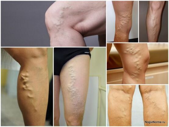
Typically, varicose veins look like an expansion of veins, which causes cosmetic inconvenience, causes pain in the legs, cramps at night, trophic changes in the area of the legs.The expansion of the veins are different-from small vascular stars and knots to large stem trunks, protruding plexuses, which are clearly visible in an upright position.
In 75 - 80 % of cases, the barrel or branches of a large subcutaneous vein suffer.Lesser Vienna is easier - its lesions are observed only in 5 - 10 % of cases.When feeling the vein seems to be elastic, it can be easily compressed, and the temperature over the affected vessel is slightly higher than in all other parts of the epidermis.This is due to the release of arterial blood and blood from deep veins through the communicative vessels to varicose nodes located on the surface.In a horizontal position, the voltage with veins subsides, and the dimensions of the affected nodes are reduced.In some cases, it is possible to probe small defects in the places of compounds of perforant and surface veins.The longer the disease develops, the more unpleasant symptoms are felt.Fast fatigue, a feeling of heaviness in the lower extremities, paresthesia, swelling on the legs and feet appear.The latter usually arise closer in the evening, and in the morning after rest they disappear.
What does varicose veins look like - photos and signs
When a woman begins to notice even insignificant vascular nets on the surface of the skin of the lower extremities, she has the question of what varicose veins look on her legs.The disease belongs to insidious, as they are able to not manifest for a large amount of time.
Description of the disease
Varicosis of the lower extremities is stagnation in the blood system that causes the expansion of veins.In 80% of cases, the disease occurs precisely on the legs, since they account for the largest load due to walking, constant physical activity.
Each vein on the leg has a specific valve responsible for the correct blood flow.This helps to avoid stagnation of blood and a malfunction of the circulatory system.The disease occurs when the blood flow is disturbed, the blood begins to flow not only up, but also down.This happens due to improper operation of the venous valves.The vessels lose their former elasticity, deform.This is the initial stage of varicose veins.
Varicose veins can appear on any part of the legs: on the hips, under the knee, on the lower leg, on the feet.Varicosis on the legs is manifested in the form of blue stars, small red or blue patterns.
Vascular surgeons warn that the risk zones of varicose lesions are hips and lower legs.A woman should be careful about the state of the legs and timely notice the changes that arise.
On the hips, the disease manifests itself in the form of large lines resembling rivers.In the photo of varicose veins on the legs, vascular nets, stars are noted.On the lower leg, the structure of pathology is similar to the location on the hips, but only at the initial stage.With the neglect of the disease, large bruises appear on the lower leg, a rash that resembles blue and red bags.
Noticing the disease is most difficult under the knee.There are small discharge of the veins of a blue shade, red nets can appear.Varicosis on the feet is the most dangerous of all locations, the site is more susceptible to negative influences of external factors, physical activity.
At the first signs presented in the photo of varicose veins on the legs (small red nets, cobwebs, blue dots), it is necessary to purchase elastic stockings, consult a vascular surgeon.
Degree of defeat
The disease is classified according to the degree of vascular damage and veins on the legs.Modern doctors use the international classification to make a diagnosis.It distinguishes 7 main degrees of varicose veins on the legs:
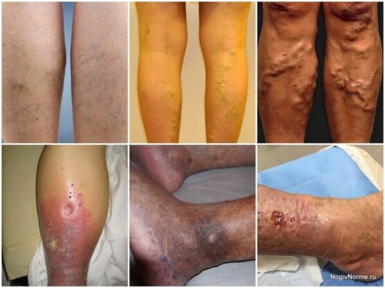
- Zero, difficult to diagnose.It is impossible to identify, it is impossible to notice the signs of the disease.Many women write them off on the usual fatigue of the legs.There is a severity in the legs, “buzz”, swelling, rare convulsions (especially at night).If you conduct an examination, it can give a false negative result;
- 1 degree looks in the photo, like small nets and stars on the legs.The patient also feels all the accompanying signs from the zero degree.They appear closer in the evening or slightly during the day;
- 2 degree - more pronounced by symptoms, a person can independently recognize the disease.Blue and red lines occur on the surface of the legs, stars and mesh acquire a bulging look.The patient begins to notice that the veins have become much larger.Signs of pathology become more pronounced after the active load on the limbs; the higher the degree of damage to the legs, the more likely to obtain complications on the cardiovascular system.Varicosis is a disease of the entire circulatory system, therefore it is accompanied by pathologies in cardiac activity, impaired blood formation process.
Types of the disease
Varicosis can be damaged by certain vessels.Depending on which of them they failed in work, doctors distinguish several types of pathology:
To understand what the disease looks like, it is necessary to consider each type of varicose veins on the legs separately:
- The name of the reticular comes from reticular veins, having a diameter of the size of not more than 2 mm.This species will be presented in the photo in the form of small nets, thin lines, stars on the legs.They are noticeable, as they are located near the surface of the dermis.Women often notice such manifestations on the side surface of the thigh.Reticular varicose veins is not dangerous.Such a variety of the disease does not bring discomfort (except for aesthetic), it does not cause serious complications.
- Magistral appearance - the trunk of a small or large subcutaneous vein is affected, which leads to the expansion of protocol veins.The name also comes from a failure in the work of a certain vein - trunk.If a large vessel is damaged, the picture of varicose veins on the legs will be this: large tubercles similar to rivers, curved lines.When malfunctioning in the work of a small vein, similar signs occur, but having a less pronounced form.This is a dangerous form of pathology, which threatens with untimely treatment with serious complications;
- Segmental.The prototing veins are affected, with a diameter of 3 mm.These are large vessels, so the disease can be detected independently.Symptoms are presented in the photo of varicose veins in the initial stage of the segmental type.It represents small swelling, small tubercles on the surface of the dermis.They do not have a blue or red shade.With untimely seeking a doctor, tubercles can expand and turn into large bruises.In case of early diagnosis from the disease, you can get rid of sclerotherapy;
- Perforant and most dangerous varicose veins, comes from the name of perforant veins.They play a huge role in the circulatory system.These veins connect deep vessels to surfaces.Often such a defeat leads to surgery.In the photo, the disease resembles large or medium -sized tubercles, irregularities on the surface of the skin.They can be single or merge into a single structure, becoming even more voluminous in size.
It is important to seek medical help in order to avoid unpleasant consequences, not to bring the disease to the need for surgery.The main one is also the prevention of varicose veins.It consists in wearing convenient correct shoes made of natural fabrics with orthopedic sole.Do not give an excessive load on the lower limbs.Men, like women should be more attentive to their feet, often inspect them on the presence of damage and symptoms of varicose veins.
What does varicose veins look like - photos and signs
When a woman begins to notice even insignificant vascular nets on the surface of the skin of the lower extremities, she has the question of what varicose veins look on her legs.The disease belongs to insidious, as they are able to not manifest for a large amount of time.
What does varicose veins look like?
Varicose veins - the disease is very insidious, since the first signs of its development usually pass for a person imperceptibly.As a rule, the patient perceives fatigue and heaviness in the legs as a consequence of heavy workdays.
In many cases, it is, but this is not a reason not to monitor the state of blood vessels on the lower extremities.Therefore, we will further talk about what this disease is, and also consider the types and degree of manifestation of varicose veins in the photo.
Varicose veins - characteristics of pathology
Varicosis on the legs is not only a cosmetic problem that causes people's concern about their appearance, but also a very dangerous pathology that requires adequate, timely therapy and observance of preventive measures.
Reference.Famine faces suffer from varicose veins several times more often than men.
Varicose veins of the lower extremities- A disease that is characterized by the emergence of the expansion of veins.In addition, there is a violation of the blood outflow, which leads to stagnant phenomena in the circulatory system.
In order to understand what varicose veins are (see photo), you should consider the process of its occurrence.
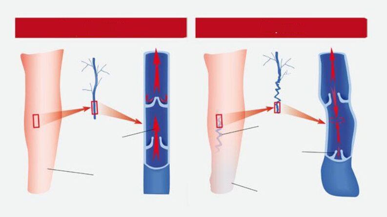
Veins located on the legs havespecial valves.They carry outBlood pass exclusively in one direction (up) and prevent it from stagnaning.
The failure in the functioning of these valves leads to the fact thatBlood gets the opportunity to move in the opposite direction (down), which prevents its outflow.
As a result, the vessels lose normal elasticity and under blood pressure begins to expand and deform.All this provokes the development of the initial stage of the disease.
Reference.Symptoms of the disease can manifest in various areas of the lower extremities, especially on the hips, under the knees, on calves, varicose veins are also often found (photo below).
Factors provoking the development of this pathology are a huge number:Starting with a sedentary lifestyle, physical overloads, bad habits and ending with hereditary predisposition, hormonal malfunctions, and congenital weakness of the walls of blood vessels.
Many people, without realizing the seriousness of the problem, are in no hurry to contact a specialist, but begin to practice self -medication.
However, this is not an output, since the independent use of vascular drugs is harmful to health.In addition, they are not able to solve the problem.A competent integrated approach is needed here.
Having determined the features of the development of pathology, then we will consider how varicose veins on the legs look depending on the type of development.
The degree of varicose veins
The community of scientists of the world systematized the manifestations of varicose disease of the lower extremities.
Reference.This classification is called Clinical Etiology Anatomy Pathology).
In accordance with the compiled classification, 7 degrees (stages) of the disease are distinguished:
- zero- The condition in which a person experiences severity in the legs, swelling, convulsions, but not manifested visual.During special examinations, no deviations are determined;
- 1 degree varicose veins (see photo below)- vascular stars appear and the same signs are observed as in the previous degree;
- The second- the bulging veins or nodules of blue color become noticeable, which can increase in size from excessive physical exertion or prolonged stay in one position;
- The third- All of the above features are supplemented by regular swelling, which is especially noticeable in the evening.In the morning, it can disappear, but by the evening it appears again;
- Fourth-Due to power failure, trophic processes in the vascular system begin.Around the veins, the skin becomes brown or black.Inflammation on the skin may develop: from small redness to large weeping wounds;
- Fifth- all symptoms of the 4th degree plus pronounced prolonged trophic ulcer;
- Sixth- Actively acting, non -healing trophic ulcer.
Such a disease should be treated immediately, and the better the better.The last stages of varicose veins are dangerous, but also possible serious complications that can lead to death.
In addition, photos of the first stage of varicose veins look quite normal, but the photos of the subsequent are shocked.
Conclusion
Information and pictures of varicose veins should make you think about what you should be attentive to your health and lead the right lifestyle.Preventive measures in this case are the ability to avoid this disease.
If, by virtue of the anatomical characteristics of the body, vein defeats occurred, then they should immediately be treated.
Varicose veins need urgent treatment, the absence of which can turn into serious consequences: the development of thrombophlebitis, bleeding from venous nodes and a trophic ulcer, capable of provoking amputation of the leg.